
Root Canal Treatment: Saving a Severely Infected Molar and Restoring Pain-Free Bite Function
PATIENT DETAILS
Age: 38 | Gender: Female
Medical History: No systemic diseases, no known drug allergies. Non-smoker, non-diabetic, not on any long-term medication. The patient had avoided dental visits for approximately three years and presented with acute dental pain. She reported a history of a large cavity on the lower left first molar (tooth 36) that had been filled once at a local clinic but continued to deteriorate. A 38-year-old homemaker, apprehensive about dental procedures but motivated to save her tooth rather than proceed with extraction.
CASE PRESENTATION
A 38-year-old female patient presented at Impladent® Dental Clinic in Malad West, Mumbai, with the chief complaint of severe, spontaneous throbbing pain in the lower left jaw that had been worsening over five days. The pain was exacerbated by hot foods and lingered long after the stimulus was removed. She reported disturbed sleep on two consecutive nights and the inability to chew on the affected side. She had been managing the pain with over-the-counter analgesics, which provided only partial and temporary relief.
On clinical examination, tooth 36 demonstrated sharp pain on percussion and prolonged sensitivity to heat. The tooth had a deep existing composite restoration with secondary caries visible at the margins on periapical radiography. The periapical X-ray revealed a periapical radiolucency of approximately 4 mm in diameter at the mesial root apex, consistent with a periapical abscess. Pulp vitality testing produced no response, confirming pulp necrosis. There was no evidence of vertical root fracture, external resorption, or furcal involvement. Probing depths were within normal limits at all sites.
The patient was evaluated by Dr. Glenn Mascarenhas, senior prosthodontist at Impladent®. Following clinical and radiographic assessment, she was confirmed as a suitable candidate for root canal treatment of tooth 36 with subsequent full-coverage crown restoration. The treatment plan was explained in detail, including the number of appointments, the procedure involved, and the importance of timely crown placement to prevent post-treatment fracture.
Root Canal Treatment Journey: From Acute Infection to a Fully Restored Tooth
STEP 1 Initial Consultation | STEP 2 Clinical & X-Ray Exam | STEP 3 Access & Pulp Removal | STEP 4 Canal Shaping & Irrigation | STEP 5 MTA Obturation | STEP 6 Temporary Seal & Review | STEP 7 Crown Placement |
| Patient reported severe throbbing pain and hot sensitivity at tooth 36 lasting five days | Periapical X-ray confirmed periapical abscess; pulp vitality test confirmed pulp necrosis | Access cavity prepared under IANB; necrotic pulp tissue removed from all three root canals | Rotary NiTi files used; NaOCl and EDTA irrigation with ultrasonic activation to clear bacteria | MTA apical plug placed at mesial canals; warm vertical compaction with gutta-percha and AH Plus | Cavity sealed with Cavit; patient reviewed at seven days confirming full pain resolution | Full-coverage metal-ceramic crown placed; bilateral chewing and normal bite fully restored |
“Root canal treatment is not a procedure to fear; it is a procedure that relieves the very pain patients associate with it. The goal is to eliminate the source of infection, seal the canal system, and restore the tooth to full function with a protective crown.” Dr. Glenn Mascarenhas, Impladent® Dental Clinic, Malad West, Mumbai |
The root canal treatment patient journey: diagnosis, multi-canal endodontic therapy, MTA obturation, and restored chewing function with a full-coverage crown.
DIAGNOSIS
Irreversible pulpitis progressing to pulp necrosis with chronic periapical abscess at tooth 36 (lower left first molar). Three-canal molar with periapical radiolucency at mesial root apex measuring approximately 4 mm. No periodontal disease, no vertical root fracture, no furcal bone loss. ASA I classification. Confirmed candidate for non-surgical root canal treatment under local anaesthesia followed by full-coverage metal-ceramic crown.
Irreversible pulpitis is a condition in which the dental pulp (the soft tissue inside the tooth containing nerves and blood vessels) becomes severely and irreversibly inflamed, usually as a result of deep bacterial invasion from untreated caries. Once the pulp becomes necrotic, bacteria and their by-products extend through the root apex into the surrounding periapical bone, forming an abscess. Without endodontic intervention this infection cannot resolve and may spread. Root canal treatment removes the infected tissue, disinfects the canal system, and seals it to prevent reinfection, allowing the natural tooth to be retained and restored to normal function.
TREATMENT
- Pre-operative Assessment: Clinical examination, periapical and bitewing radiography, pulp vitality testing, patient medical and dental history review
- Emergency Pain Management: Inferior alveolar nerve block (IANB) administered; drainage of periapical abscess confirmed via access cavity; initial debridement performed at first visit
- Canal Preparation: Working length established using an apex locator and confirmed with periapical X-ray; three canals (mesiobuccal, mesiolingual, distal) shaped using rotary NiTi files
- Canal Irrigation and Disinfection: Sequential irrigation with 2.5% sodium hypochlorite (NaOCl), 17% EDTA, and normal saline; passive ultrasonic activation to improve disinfection efficacy
- MTA Apical Plug and Obturation: Mineral trioxide aggregate (MTA) placed at the apical third of the mesial canals; warm vertical compaction with gutta-percha and AH Plus sealer for final obturation
- Temporary Restoration and Review: Access cavity sealed with Cavit; patient reviewed at seven days for pain resolution confirmation
- Full-Coverage Crown Restoration: Tooth 36 restored with a metal-ceramic crown for long-term structural protection
DETAILED DESCRIPTION OF TREATMENT
Following the confirmed diagnosis of pulp necrosis with periapical abscess, Dr. Glenn Mascarenhas initiated the root canal treatment protocol across two primary clinical appointments. The decision to use MTA as an apical barrier material was based on the periapical radiolucency at the mesial root, which indicated apical breakdown requiring biocompatible sealing rather than a standard gutta-percha condensation at the terminus.
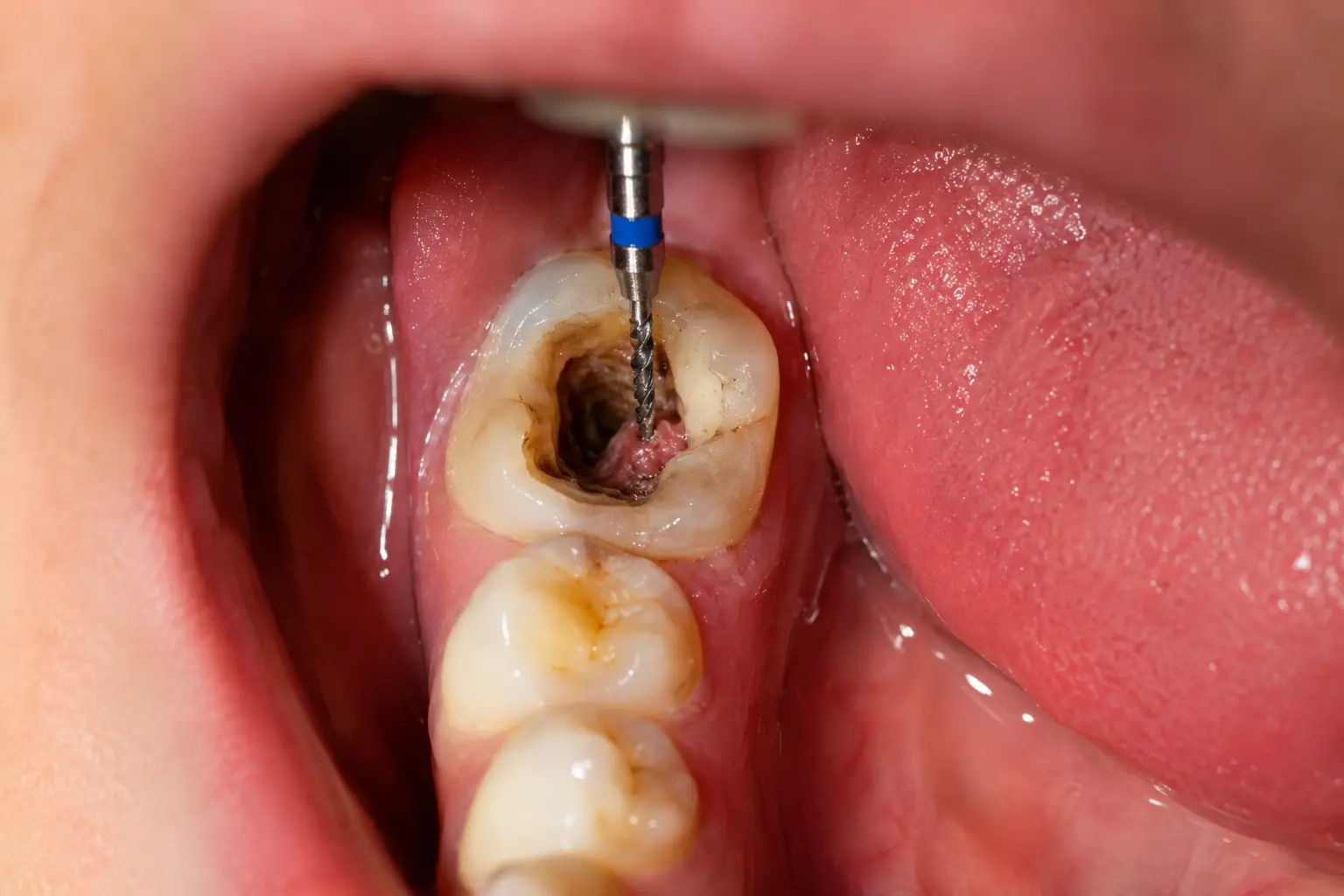
The first appointment began with administration of an inferior alveolar nerve block (IANB) on the left side. Given the patient’s anxiety, a slow-speed injection technique was used with a buffered lignocaine formulation, and the patient was reassured throughout. Access cavity preparation was performed through the existing composite restoration using a diamond bur to create a triangular outline form appropriate for a mandibular molar. On opening, purulent exudate was visible in the pulp chamber, confirming the periapical abscess. This was suctioned, and the chamber irrigated immediately with 2.5% NaOCl.
Working lengths for all three canals (mesiobuccal MB, mesiolingual ML, and distal D) were established using an electronic apex locator and verified with a periapical radiograph. Canal shaping was performed with a sequence of rotary NiTi files, with alternating NaOCl and 17% EDTA irrigation and a final passive ultrasonic activation step. The canals were dried with sterile paper points. MTA was mixed to a putty consistency and placed at the apical 4 mm of the MB and ML canals; correct positioning was verified radiographically. A moist cotton pellet was placed over the MTA and the access cavity was sealed with Cavit. The patient was prescribed amoxicillin 500 mg three times daily for five days and ibuprofen 400 mg for pain. For complete information on the procedure, candidacy, and cost, visit our root canal treatment in Malad, Mumbai page.
“MTA is our preferred apical material in cases with established periapical pathology. Its biocompatibility and ability to set in the presence of moisture make it particularly suitable for cases with apical lesions, where complete dryness of the canal terminus is difficult to guarantee.”
Dr. Glenn Mascarenhas, Senior Prosthodontist, Impladent® Dental Clinic
At the second appointment seven days later, the patient reported complete resolution of pain and the absence of swelling. The Cavit was removed, the MTA was verified as set, and warm vertical compaction of the distal canal and the coronal portion of the mesial canals was performed using gutta-percha and AH Plus sealer. A post-obturation periapical radiograph confirmed three-dimensional filling of the root canal system with no voids, overfills, or missed canals. The access cavity was cleaned and filled with a glass ionomer base, and a full metal-ceramic crown was placed at a dedicated restorative appointment. Crown occlusion was adjusted to eliminate premature contacts in centric and lateral excursions.
POST-OPERATIVE ASSESSMENT
The patient was reviewed ten days after crown placement. She reported no pain, sensitivity, or swelling. Chewing on the left side was fully restored and comfortable. She confirmed she had resumed a normal diet including firm foods without discomfort. Clinical examination showed a well-adapted crown margin with no tenderness on percussion or palpation at the periapical area.
“I honestly could not believe how painless the treatment was. I had been scared for nothing. The moment the infection was cleared, the pain simply went away, and now I eat normally without even thinking about it.” Patient |
At the six-month follow-up, a periapical radiograph demonstrated significant resolution of the periapical radiolucency at the mesial root. The lesion had reduced from approximately 4 mm to a near-normal trabecular pattern around the apex, indicating bone regeneration consistent with successful endodontic healing. The patient remains on a six-month clinical recall and annual radiographic review protocol. No signs of reinfection, periapical pathology, or crown failure were observed at the time of the six-month review. Patients with multiple teeth requiring restoration following endodontic treatment may also be candidates for full mouth rehabilitation, which addresses comprehensive occlusal and aesthetic restoration in a structured treatment sequence.
This case illustrates a fundamental principle in conservative dentistry: every effort should be made to retain the natural tooth before considering extraction. Root canal treatment, when performed with precision (correct working length establishment, thorough canal disinfection, and appropriate obturation), predictably eliminates infection and allows periapical healing. The subsequent full-coverage crown is not optional; it is an essential component of the treatment that protects the endodontically weakened tooth from fracture and ensures long-term function.
CONTACT — IMPLADENT® DENTAL CLINIC, MALAD WEST, MUMBAI
Treating Dentist: | Dr. Glenn Mascarenhas, Senior Prosthodontist & Implantologist |
Co-Specialist: | Dr. Sonia Butta, Prosthodontist |
Clinic: | Premier Tower, 201/301, Marve Road, Orlem, Malad West, Mumbai, Maharashtra 400064 |
Phone: | +91 022 31912633 | +91 022 31424745 |
Website: |
Impladent® is a premium dental clinic in Malad, delivering advanced dental solutions with a commitment to precision, safety, and excellence in patient care.